Charting a safe course
All forms of brain surgery require an extraordinary level of planning and skill to reduce the risk of harming sensitive regions of the brain that control essential bodily functions, such as the motor cortex, which governs muscle movement. The same is true of back surgery, which requires surgeons to steer clear of the spinal cord and nerves exiting the spine when repairing diseased or injured discs and vertebrae. Damage to critical brain tissue or nerves can lead to complications such as limb weakness or numbness, to paralysis or loss of speech or vision.
To minimize such risks while taking out a brain tumor or treating a spinal disorder, the goal is straightforward, said Gordon Nakata, MD, FAANS, a neurological surgeon at Cape Cod Hospital: “Take the safest path possible.”
To chart a safe course when removing a tumor from inside the brain, a key pre-planning step is identifying its location in relation to regions that manage movement or control the senses, said his fellow neurological surgeon Nicholas Coppa, MD.
“We know roughly where (these regions) are laid out in brain,” explained Dr. Coppa. So, if an MRI indicates that a tumor is next to the motor cortex, “that’s where I need to be careful.”
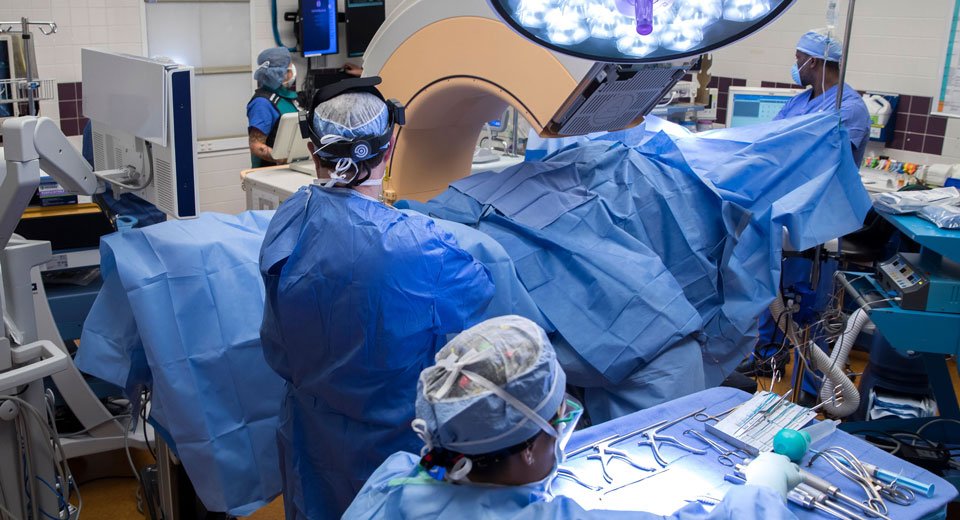
To augment their knowledge of the brain’s anatomy, neurological surgeons at Cape Cod Hospital turn to image-guidance technology called the StealthStation Surgical Navigation System (which is also used with the O-arm Imaging System. During the procedure, this system synchronizes the MRI of the brain taken before the operation with real-time images captured by an infra-red camera. This allows the surgeon to track the movement of the very small scalpels and other tiny instruments used to remove a brain tumor. With the aid of the detailed, three-dimensional images provided by the StealthStation technology, “we can get the shortest, safest trajectory to the tumor,” said Achilles Papavasiliou, MD, FAANs, a neurological surgeon at Cape Cod Hospital.
Other technology, such as electromyography—which measures muscles’ response to signals from the brain—is often also used in neurological surgery to guard against nerve damage. Electrodes placed in the legs and arms of a patient undergoing brain or back surgery transmit information about nerve signals reaching the muscles in these limbs.
A doctor called a neurophysiologist monitors these signals on a computer screen during an operation. So, for example, if a screw implanted into a vertebra during a disc fusion procedure irritates or compresses a nerve, the screen flashes a warning and the neurophysiologist alerts the surgeon. In such a case, a smaller screw might be necessary. In the event a surgeon irritates a pinched nerve during the removal of bone or disc, “simply slowing down until the stimulation resolves is all that is necessary,” said Dr. Papavasiliou. In other cases, the danger of harming a nerve may require the surgeon to use smaller instruments or approach the tissue that needs to be excised or repaired from another angle.
Known as neuromonitoring, the various technologies used to keep tabs on whether nerves are in any danger during surgery give doctors immediate feedback that helps prevent post-operative side effects and complications. Coupled with advances in imaging technology, they add up to better surgical outcomes and greater safety for patients.
